
At a glance
-
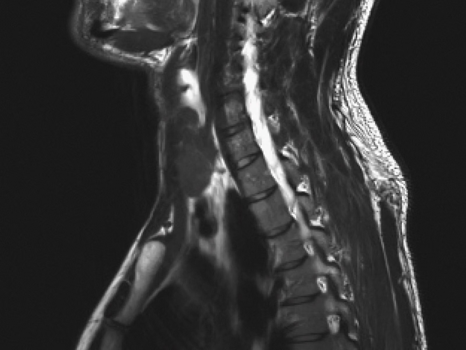
The MRI with Spine includes everything in Function’s Annual MRI, plus targeted imaging of your spine, helping identify signs of spinal lesions and tumors, herniated discs, cervical spinal stenosis, spondylosis, fractures, scoliosis, arthritis, and other degenerative changes.
Your spine does a lot of work. It holds you upright, protects the nerves that connect your brain to the rest of your body, and absorbs the impact of everything from a morning run to sitting at a desk all day. But most people don't think about their spine until something hurts.
Function’s MRI with Spine gives you a closer look at your spine as part of your annual health check.
Why Function offers MRI spine coverage
Function's Annual MRI already covers your head, neck, abdomen, and pelvis to look for signs of cancer (brain, liver, pancreas, kidneys, ovaries, prostate, and more), prior stroke, aneurysms, fatty liver, and hundreds of other conditions. The MRI with Spine adds a dedicated look at your spine on top of that.
A doctor might only order a spine MRI once you already have certain symptoms such as numbness or tingling, or pain after an injury. These diagnostic MRIs include multiple views to identify specific issues. But spine problems often develop slowly and without obvious symptoms until they've progressed. That’s why Function includes coverage with a key view of the spine as an option so you can look for signs of many conditions early, rather than waiting for symptoms to show up.
What the MRI with Spine can find
The MRI with Spine includes everything in the Annual MRI, plus targeted spine coverage that helps identify signs of:
- Spinal lesions and tumors: Can form anywhere along the spine and, depending on location, may press on nearby nerves or the spinal cord.
- Herniated discs: Where disc material pushes through a tear in the tough outer layer of a disc and can irritate nearby nerves.
- Cervical spinal stenosis: A narrowing of the spinal canal in your neck that can compress the spinal cord or nerve roots and lead to numbness, weakness, neck pain, or neurologic symptoms in the arms or legs.
- Spondylosis: A form of arthritis affecting the spine that develops as discs and joints wear down over time, often without any symptoms in its early stages.
- Fractures: Include compression fractures that can result from trauma, osteoporosis, or a tumor weakening the vertebra.
- Scoliosis: An abnormal sideways curve in the spine that can affect posture, mobility, and in some cases, lung function.
- Arthritis throughout the spine's joints: Can restrict range of motion and contribute to chronic stiffness.
- Other degenerative changes: In the discs and joints of your spine, such as bone spurs or disc dehydration, that build up gradually with age and repeated strain.
These findings can affect different parts of your spine in different ways.
A closer look at your spine's anatomy
Your spine is made up of 33 vertebrae stacked from the base of your skull to your tailbone. Discs and joints connect them, helping you bend, twist, and absorb impact.
Your spinal cord runs through the upper portion of your spine before giving way to a bundle of nerve roots called the cauda equina lower down. Together, these nerves carry signals between your brain and the rest of your body.
The MRI with Spine looks at several regions, each with a different role.
The cervical spine
Your cervical spine, or neck, has 7 vertebrae and 8 nerve branches. It supports your head, allows you to turn and tilt your neck, and protects the arteries that carry blood to your brain.
An MRI here can help identify signs of:
- Cervical spinal stenosis, the narrowing of the spinal canal, which can compress the spinal cord and nerves
- Herniated discs
- Bone spurs
- Cervical degenerative disc disease, the gradual wearing down of discs over time
- Cervical spondylosis, a form of arthritis in the neck
The thoracic spine
Your thoracic spine is the longest section, running between the base of your neck and middle back. It has 12 vertebrae and 12 nerve branches, and it anchors your rib cage while protecting nerves that connect to your lungs, heart, and liver.
An MRI here can help identify signs of:
- Spinal tumors
- Bulging discs
- Bone spurs
- Compression fractures, where a vertebra collapses, often due to trauma, osteoporosis, or a tumor
The lumbar spine
Your lumbar spine (lower back) has 5 vertebrae and 5 nerve branches, and it carries most of your body weight.
An MRI here can help identify signs of:
- Herniated discs
- Lumbar stenosis
- Spondylolisthesis (a slipped vertebra)
- Compression fractures
- Nerve root compression
- Unusual curvature such as scoliosis
Why you might want spine coverage
Back and neck problems are common, and they tend to build gradually rather than show up overnight. Adding spine coverage to your MRI gives you a way to check for changes such as arthritis, disc degeneration, and early curvature changes before they might turn into the kind of pain that sends many people to a doctor in the first place.
You might also want to add spine coverage if you spend long hours sitting at a desk, work a physically demanding job, or have a family history of arthritis, osteoporosis, or scoliosis. Athletes and people who lift heavy weights regularly also put repeated strain on their discs and joints, which can accelerate wear over time. And with age, spine issues tend to become more common, since discs naturally lose hydration and joints experience more wear with each decade.
Knowing what’s happening in your spine gives you information you can use—whether that means discussing a finding with your doctor, working with a physical therapist, changing how you move or sit, or simply keeping an eye on something over time.
Get a clearer view of your health with Function
Function’s 160+ yearly lab tests show what’s circulating in your blood and urine—inflammation, heart health, metabolism, nutrients, and more. MRIs*, including the MRI with Spine, show what’s inside your body—organs, tissues, and other changes. That’s why Function recommends getting both every year for a clearer picture of your health that you can track over time. *Available for an additional cost.
End notes
- Expert Panel on Neurological Imaging, Hutchins TA, Peckham M, et al. ACR Appropriateness Criteria® Low Back Pain: 2021 Update. J Am Coll Radiol. 2021;18(11S):S361-S379. doi:10.1016/j.jacr.2021.08.002
- Kumar N, Tan WLB, Wei W, Vellayappan BA. An overview of the tumors affecting the spine—inside to out. Neurooncol Pract. 2020;7(Suppl 1):i10-i17. doi:10.1093/nop/npaa049
- Scarcia L, Pileggi M, Camilli A, et al. Degenerative Disc Disease of the Spine: From Anatomy to Pathophysiology and Radiological Appearance, with Morphological and Functional Considerations. Journal of Personalized Medicine. 2022;12(11):1810. doi:10.3390/jpm12111810
- Baradaran Mahdavi S, Riahi R, Vahdatpour B, Kelishadi R. Association between sedentary behavior and low back pain; A systematic review and meta-analysis. Health Promot Perspect. 2021;11(4):393-410. doi:10.34172/hpp.2021.50
- Johnson MA, Gohel S, Mitchell SL, Flynn JJM, Baldwin KD. Entire-spine Magnetic Resonance Imaging Findings and Costs in Children With Presumed Adolescent Idiopathic Scoliosis. J Pediatr Orthop. 2021;41(10):585-590. doi:10.1097/BPO.0000000000001943
- Anaya JEC, Coelho SRN, Taneja AK, Cardoso FN, Skaf AY, Aihara AY. Differential Diagnosis of Facet Joint Disorders. RadioGraphics. 2021;41(2):543-558. doi:10.1148/rg.2021200079
- Yang D, Liu C, Hu Q. Diagnostic value of a magnetic resonance imaging (MRI)-based vertebral bone quality score for bone mineral density assessment: an updated systematic review and meta-analysis. Quant Imaging Med Surg. 2024;14(10):7561-7575. doi:10.21037/qims-24-532
- Salo S, Hurri H, Rikkonen T, Sund R, Kröger H, Sirola J. Association between severe lumbar disc degeneration and self‐reported occupational physical loading. J Occup Health. 2022;64(1):e12316. doi:10.1002/1348-9585.12316
- Ekşi MŞ, Orhun Ö, Yaşar AH, et al. At what speed does spinal degeneration gear up?: Aging Paradigm in patients with Low Back Pain. Clinical Neurology and Neurosurgery. 2022;215:107187. doi:10.1016/j.clineuro.2022.107187